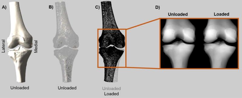
Fig. 2.

An overview of the method for creating the mimicked fluoroscopy images. A) Bones segmented from the CT scan are loaded into MATLAB and positioned based on kinematic data. In this case, the unloaded bone is shown. B) Based on the outline of the bone, a regular matrix of binary data points was created that identified which points fell within the bone. Here, the points on the edge of that point cloud are shown overlaid onto the bone. C) Comparison of point clouds (edges only) in the unloaded and loaded states. The perfectly aligned femurs can be seen. D) Mimicked fluoroscopy (mFluoro) images were created by projecting these point clouds onto a 2D image.
