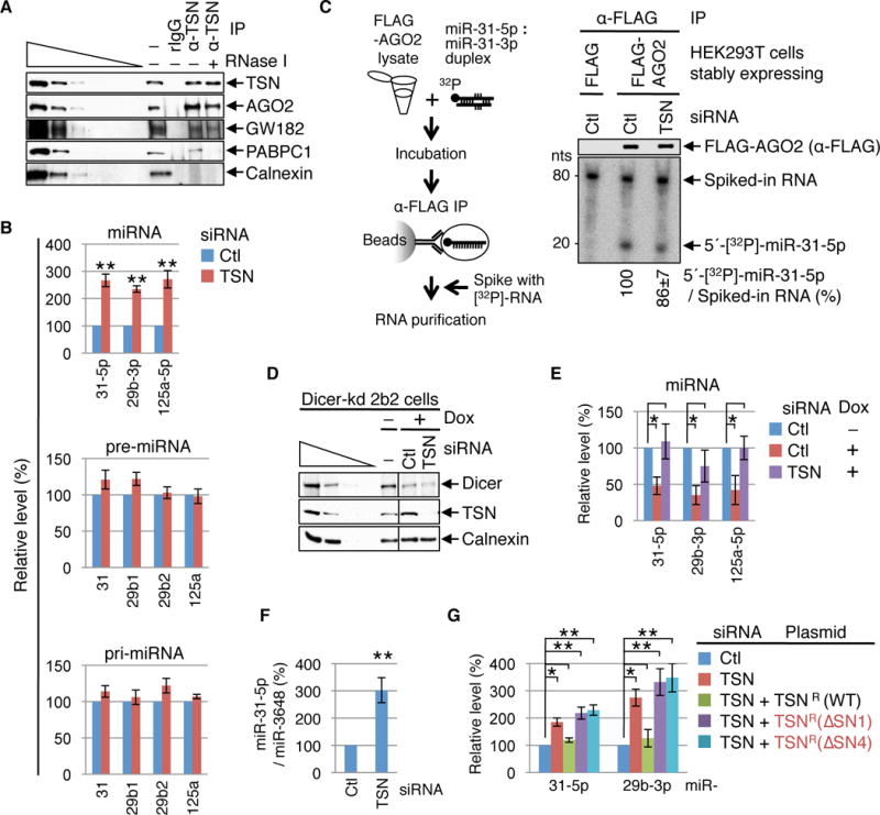
Fig. 1. TSN mediates cellular degradation of mature and biologically active miRNAs.

(A) Western blot (WB) demonstrating that HEK293T-cell TSN co-immunoprecipitates with RISC components in the presence (+) or absence (−) of RNase I. RNase I activity is confirmed by the loss of PABPC1 from the TSN IP, and IP specificity is evident by the absence of Calnexin. The wedge specifies three-fold dilutions of cell lysates. rIgG, control rabbit IgG; α-TSN, anti-TSN; − IP, prior to IP.
(B) RT-qPCR revealing that mature miRNA levels increased in cells transfected with TSN siRNA relative to Ctl siRNA, while the corresponding pre- or pri-miRNAs levels remained unchanged.
(C) Left: Lysates of HEK293T cells stably expressing FLAG or FLAG-AGO2 were incubated with 5′-[32P]-labeled miR-31-5p : miR-31-3p duplexes, and α-FLAG immunoprecipitates were spiked with in vitro-synthesized, internally [32P]-labeled RNA before RNA extraction. Right: WB (upper) and phosphorimage of RNAs extracted from α-FLAG IPs (lower) showing that TSN KD does not increase the efficiency of 5′-[32P]-miR-31-5p loading into FLAG-AGO2.
(D) WB showing that exposing Dicer-kd 2b2 cells (20) to doxycycline (Dox) to induce Dicer shRNA production inhibits Dicer expression. Cells were transfected with Ctl or TSN siRNA and exposed to Dox for 4 days prior to harvesting.
(E) RT-qPCR of RNA from (D) demonstrating that when doxycycline is used to inhibit miRNA biogenesis miRNAs are stabilized by TSN siRNA relative to Ctl siRNA.
(F) RT-qPCR showing that TSN siRNA upregulates the level of exogenously introduced miR-31-5p relative to the level of co-introduced miR-3648.
(G) RT-qPCR showing that TSNR(WT), but not the catalytically inactive variant TSNR(ΔSN1) or TSNR(ΔSN4), rescues the activity of cellular TSN.
Here and elsewhere, all results derive from ≥ 3 independent experiments. For RT-qPCR results, miRNA and pre-miRNA levels are relative to U6 snRNA, pri-miRNAs levels are relative to β-actin mRNA, and relative levels in the presence of Ctl siRNA are defined as 100. Histograms represent the average and SD. *P < 0.05, **P < 0.01.
