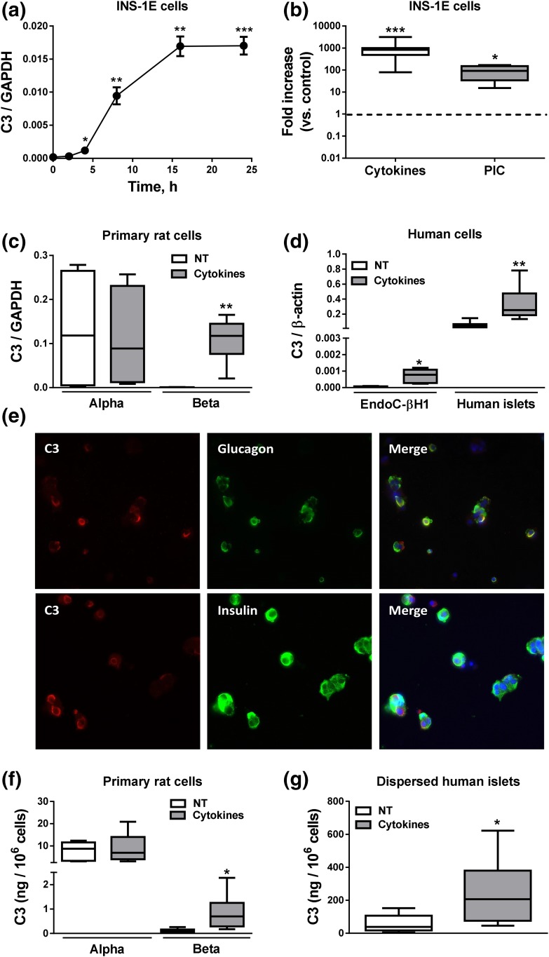
Figure 2.
Complement C3 expression in pancreatic cells. (a) INS-1E cells were left untreated or treated with IL-1β plus IFN-γ (10 and 100 U/mL, respectively) for 2, 4, 8, 16, and 24 hours. (b) INS-1E cells were left nontreated or treated with cytokines (10 and 100 U/mL, respectively) or PIC (1 μg/mL) for 24 hours. (c) Primary rat α- and β-cells were left untreated or treated with IL-1β plus IFN-γ (50 and 500 U/mL, respectively) for 48 hours. (d) EndoC-βH1 cells and dispersed human islets were left untreated or treated with IL-1β plus IFN-γ (50 and 1000 U/mL, respectively) for 48 hours. C3 mRNA expression was analyzed by real-time polymerase chain reaction (RT-PCR) and normalized by the housekeeping genes GAPDH or β-actin. (e) Immunocytochemistry of C3 (red), insulin or glucagon (green), and HO (blue) was performed to confirm C3 expression in three dispersed human islet preparations (images are representative of three independent experiments; original magnification, ×40). (f and g) C3 secretion in (f) primary rat α- and β-cells and (g) dispersed human islets was measured by ELISA. C3 secretion was normalized by number of cells. Results are means ± SEM of 4 to 23 independent experiments. *P ≤ 0.05, **P < 0.01, and ***P < 0.001 vs treated with cytokines or PIC by Student t test. NT, nontreated.

