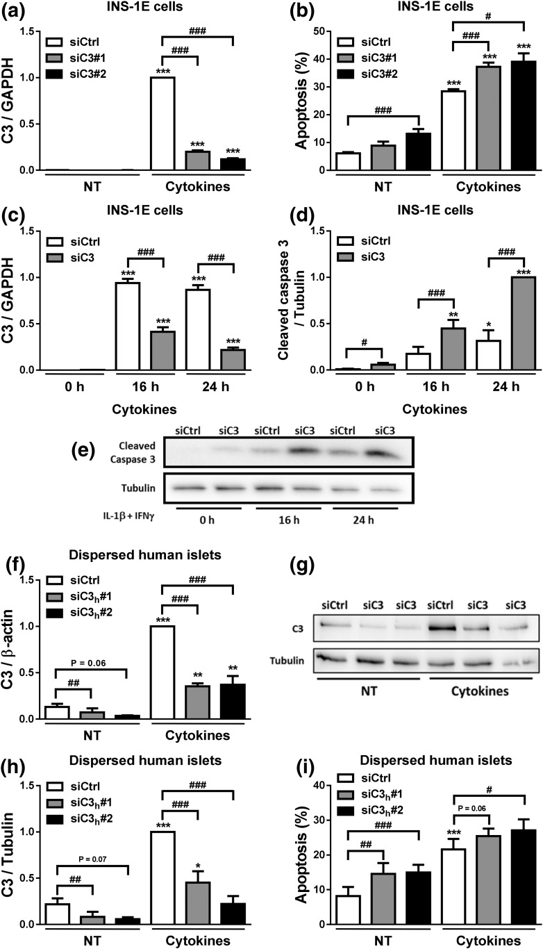
Figure 4.
C3 inhibition increases β-cell apoptosis. (a–e) INS-1E cells were transfected with siCtrl or siRNAs targeting rat C3 (siC3). Cells were then left untreated (NT) or treated with IL-1β plus IFN-γ (10 and 100 U/mL, respectively) for 24 hours. (a) C3 mRNA expression was analyzed by real-time polymerase chain reaction (RT-PCR) and normalized by the housekeeping gene GAPDH. (b) Apoptosis was evaluated using HO and PI staining in INS-1E cells. (c–e) INS-1E cells were transfected with siCtrl or siRNA targeting rat C3 (siC3 no. 1). (c) C3 mRNA expression was analyzed by RT-PCR and normalized by the housekeeping gene GAPDH. (d and e) Cleaved caspase-3 and α-tubulin were measured by western blot (images are representative of seven independent experiments). (f–i) Dispersed human islets were transfected with siCtrl or siRNA targeting human C3 (siC3h). Then, cells were left untreated or treated with IL-1β plus IFN-γ (50 and 1000 U/mL, respectively) for 48 hours. (f) C3 mRNA expression was analyzed by RT-PCR and normalized by the housekeeping gene β-actin. (g and h) C3 and α-tubulin were measured by western blot. (i) Apoptosis was evaluated using HO and PI staining in dispersed human islets. Results are means ± SEM of four to seven independent experiments. *P ≤ 0.05, **P < 0.01, and ***P < 0.001 vs nontreated with cytokines (NT) and transfected with the same siRNA; #P ≤ 0.05, ##P < 0.01, ###P < 0.001 as indicated by bars; ANOVA followed by Student t test with Bonferroni correction.

