Figure 8.
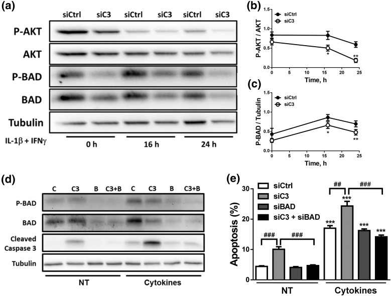
C3 silencing inhibits the AKT pathway. (a–c) INS-1E cells were transfected with siCtrl or siRNA targeting rat C3 (siC3 no. 1). Cells were then left untreated or treated with IL-1β plus IFN-γ (10 and 100 U/mL, respectively) for 16 or 24 hours. (a–c) P-BAD, BAD, P-AKT, AKT, and α-tubulin were measured by western blot. (a) Images are representative of four to seven independent experiments. (b and c) Densitometry analysis of the western blots for (b) P-AKT and (c) P-BAD. (d and e) INS-1E cells were transfected with siCtrl (C) or siRNAs targeting rat C3 (siC3 no. 1, C3), BAD (B), or both (C3 + B). Cells were then left untreated (NT) or treated with IL-1β plus IFN-γ (10 and 100 U/mL, respectively) for 16 hours. (d) P-BAD, BAD, cleaved caspase-3, and α-tubulin were measured by western blot. Images are representative of four independent experiments. (e) Apoptosis was evaluated using HO and PI staining in INS-1E cells. Results are means ± SEM of four to seven independent experiments. *P ≤ 0.05, **P < 0.01, and ***P < 0.001 vs transfected with (b and c) siCtrl vs (e) nontreated with cytokines (NT) and transfected with the same siRNA; ##P < 0.01 and ###P < 0.001 as indicated by bars; ANOVA followed by Student t test with Bonferroni correction.

