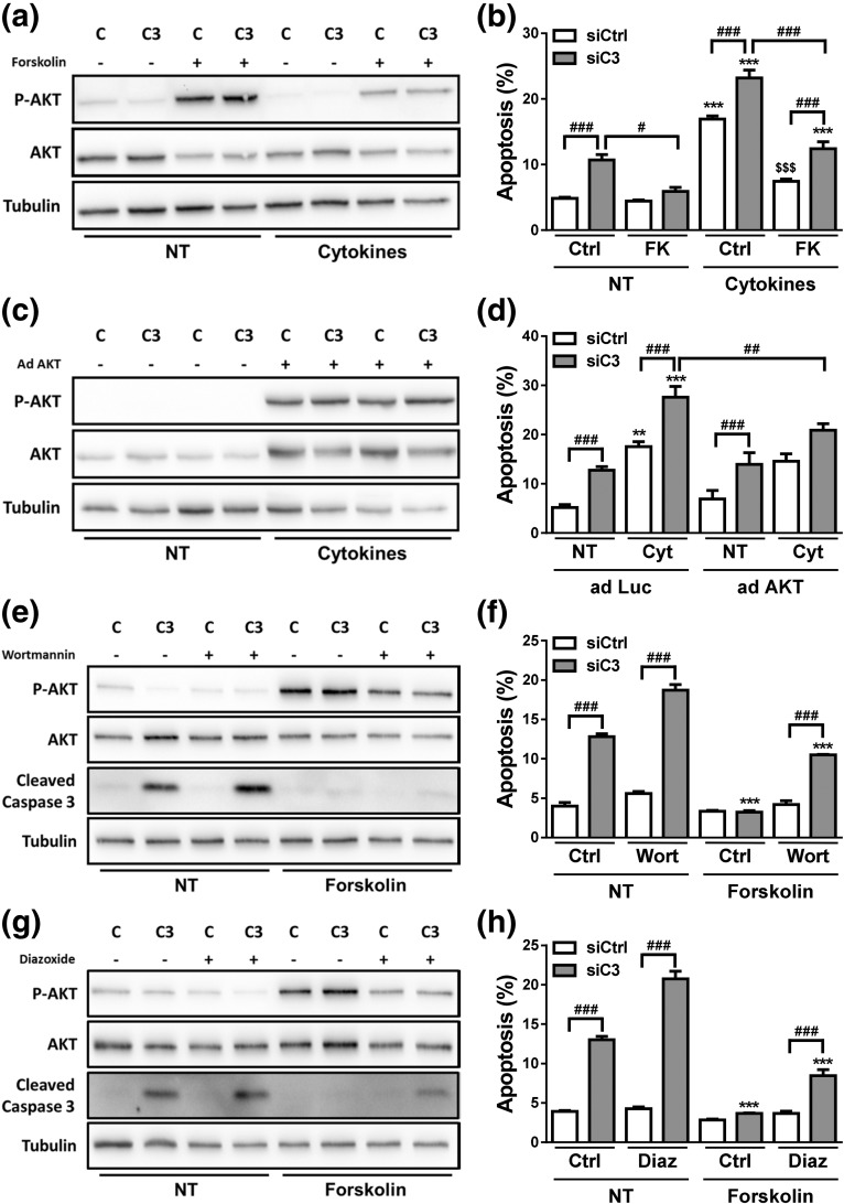
Figure 9.
AKT stimulation protects against C3 deficiency–induced apoptosis. (a and b) INS-1E cells were transfected with siCtrl (C) or siRNA targeting rat C3 (siC3 no. 1, C3). Cells were then treated with vehicle only (DMSO, Ctrl) or FK (20 µM) in the absence (NT) or presence of IL-1β plus IFN-γ (10 and 100 U/mL, respectively) for 16 hours. (a) P-AKT, AKT, and α-tubulin were measured by western blot. Images are representative of four independent experiments. (b) Apoptosis was evaluated using HO and PI staining in INS-1E cells. (c and d) siCtrl (C) and siC3 no. 1 (C3) INS-1E cells were infected with a control adenovirus encoding luciferase (adLuc) or with an adenovirus encoding myr-Akt1 (adAKT). Cells were then left untreated (NT) or treated with IL-1β plus IFN-γ (10 and 100 U/mL, respectively) for 16 hours. (c) P-AKT, AKT, and α-tubulin were measured by western blot in INS-1E cells transfected with siCtrl (C) or a siRNA targeting rat C3 (siC3 no. 1, C3). Images are representative of 4 independent experiments. (d) Apoptosis was evaluated using HO and PI staining in INS-1E cells. (e and f) INS-1E cells were transfected with siCtrl (C) or siRNA targeting rat C3 (siC3 no. 1, C3). Cells were then treated with vehicle only (DMSO, Ctrl) or wortmannin (Wort, 100 nM) in the absence (NT) or presence of FK (20 µM) for 16 hours. (e) P-AKT, AKT, cleaved caspase-3, and α-tubulin were measured by western blot. Images are representative of four independent experiments. (f) Apoptosis was evaluated using HO and PI staining in INS-1E cells. (g and h) INS-1E cells were transfected with siCtrl (C) or siRNA targeting rat C3 (siC3 no. 1, C3). Cells were then treated with vehicle only (DMSO, Ctrl) or diazoxide (Diaz, 200 µM) in the absence (NT) or presence of FK (20 µM) for 16 hours. (g) P-AKT, AKT, cleaved caspase-3, and α-tubulin were measured by western blot. Images are representative of four independent experiments. (f) Apoptosis was evaluated using HO and PI staining in INS-1E cells. Results are means ± SEM of four independent experiments. **P < 0.01 and ***P < 0.001 vs nontreated with cytokines or FK (NT) and transfected with the same siRNA; $$$P < 0.001 vs siCtrl plus cytokines; #P ≤ 0.05, ##P < 0.01, and ###P < 0.001 as indicated by bars; ANOVA followed by Student t test with Bonferroni correction.

