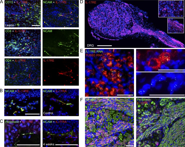
Figure 4.
Peripheral nervous systems express IL-17RE, a receptor subunit specific for IL-17c. (A) Nerve endings in lesional genital skin expressed IL-17RE. IL-17RE+ cells exhibited elongated fiber-like shapes and were distinct from CD15+, CD8a+, and CD4+ cells. Double immunofluorescent staining with anti-IL-17RE (red) and anti-CD15, CD8a or CD4 (green) antibodies revealed no costaining (left). Double immunofluorescent staining with anti-NCAM (green) and anti-IL-17RE (red) antibodies showed significant double staining of nerve endings in lesional biopsies (right). Nuclei stained with DAPI (blue). Bar, 50 µm. (B) Nerve endings in control skin biopsies express IL-17RE and IL-17RA. Double staining with anti-NCAM and anti-IL17RE or IL-17RA antibody showed double-positive nerve endings in control skin biopsies. Bar, 50 µm. (C) Nerve endings in posthealed skin biopsies express IL-17RE and IL-17RA. Double immunofluorescent staining with anti-peripherin (green) and anti-IL-17RE (red) antibodies (left) or anti-NCAM (green) and anti-IL17RA (red) antibodies (right) revealed expression of IL-17RE on peripherin+ nerve endings and expression of IL-17RA on NCAM+ nerve endings in genital 4-wk posthealed skin biopsies. Nuclei stained with DAPI (blue). Bar, 50 µm. (D) Single immunofluorescent staining with anti-IL-17RE (red) in sensory neurons from human fetal DRG showed staining in both neuronal cell bodies and nerve fibers. Insets show enlarged pictures of IL-17RE expression in cell bodies (top) and axons (bottom). Bar, 500 µm. (E) Detection of IL-17RE RNA expression in sensory neurons in human fetal DRG using FISH. TUBB3, tubulin β 3 class III. Three representative images are displayed. Bars, 50 µm. (F) IL-17RE (red) expression in a subset of NF200+ (left; green) or peripherin+ neurons (right; green) and axons in human fetal DRG. Bar, 50 µm. All the experiments were repeated three times.

