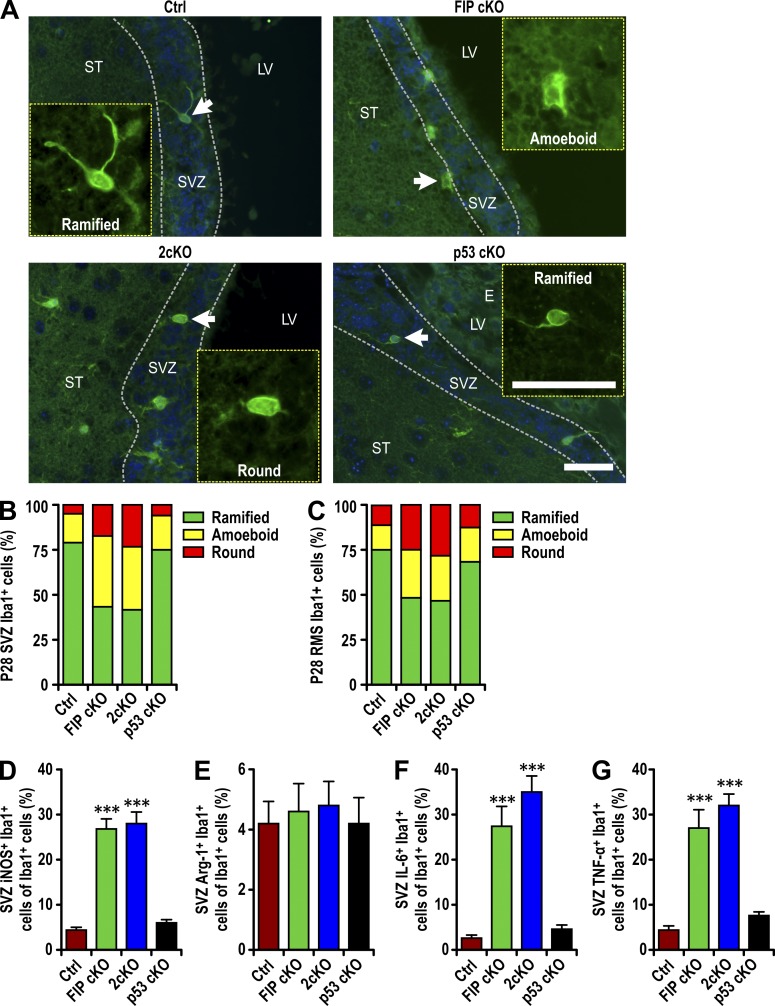
Figure 4.
Infiltrated microglia are activated in FIP200-deficient SVZ. (A) Immunofluorescence of Iba1 and DAPI in SVZ (three mice each) of Ctrl, FIP cKO, 2cKO, and p53 cKO mice at P28. Arrows indicate cells shown in more detail for Iba1 staining in insets. The dotted lines indicate the boundaries of the SVZ. Bars, 30 µm. (B and C) The percentage of ramified, round, and amoeboid microglia per SVZ (B) and RMS (C) section in Ctrl, FIP cKO, 2cKO, and p53 cKO mice at P28 (>300 cells from three mice each). (D–G) Percentage of iNOS+ microglia (Iba1+ cells; D), Arginase 1+ microglia (E), IL-6+ microglia (F), and TNF+ microglia per SVZ section (mean ± SEM; five mice each) in Ctrl, FIP cKO, 2cKO, and p53 cKO mice at P28. LV, lateral ventricle; ST, striatum; SVZ, subventricular zone. ***, P < 0.001.

