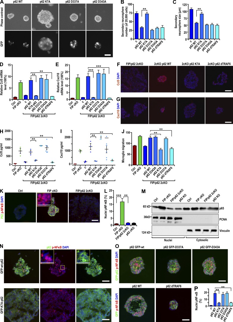
Figure 7.
Aggregated p62 promoted chemokine expression by activating NF-κB in FIP200-deficient NSCs. (A) Phase contrast (top) and GFP fluorescent (bottom) images of FIP/p62 2cKO retrovirus-infected neurospheres encoding GFP-p62-wt and its mutants as indicated. (B and C) Number (B) and the size (C) of secondary FIP/p62 2cKO retrovirus-infected neurospheres encoding GFP-p62-wt and various p62 mutations as indicated (mean ± SEM; six mice each in B and five mice each in C). (D and E) mRNA levels of Ccl5 (D) and Cxcl10 (E) in Ctrl, FIP cKO, and FIP/p62 2cKO neurospheres and FIP/p62 2cKO retrovirus-infected neurospheres encoding GFP-p62-wt and various p62 mutations as indicated (mean ± SEM; six mice each). (F and G) Immunofluorescence of Ccl5 (F) and Cxcl10 (G) with DAPI in FIP/p62 2cKO neurospheres and FIP/p62 2cKO retrovirus-infected neurospheres encoding GFP-p62-wt and its mutants as indicated (three mice each). (H and I) Concentration of Ccl5 (H) and Cxcl10 (I) in conditioned media of neurospheres (normalized to 1 mg total protein lysate of neurospheres) from Ctrl, FIP cKO, FIP/p62, and 2cKO neurospheres and FIP/p62 2cKO retrovirus-infected neurospheres encoding GFP-p62-wt and various p62 mutations as indicated (mean ± SEM; three mice each). (J) Number of migrated microglia in conditioned media from Ctrl, FIP cKO, and FIP/p62 2cKO neurospheres and FIP/p62 2cKO retrovirus-infected neurospheres encoding GFP-p62-wt and various p62 mutations as indicated (mean ± SEM; six mice each). (K) Immunofluorescence of p62 and phosphorylated p65 with DAPI in neurospheres (3threemice each) of Ctrl, FIP cKO, and FIP/p62 2cKO mice at P28. (L) Percentage of nuclei localized phosphorylated p65 from neurospheres of Ctrl, FIP cKO, FIP/p62 2cKO, and p62 KO mice are shown (five mice each). (M) Nuclear and cytoplasmic lysates extracted from neurospheres of Ctrl, FIP cKO, FIP/p62 2cKO, and p62 KO mice were analyzed by immunoblot using antibodies to detect p65, PCNA, and vinculin. (N and O) Immunofluorescence of GFP and phosphorylated p65 with DAPI in neurospheres (three mice each) of FIP/p62 2cKO retrovirus-infected mice encoding GFP-p62-wt and various p62 mutations as indicated. (P) Percentage of nuclei localized phosphorylated p65 from FIP/p62 2cKO neurospheres and FIP/p62 2cKO retrovirus-infected neurospheres encoding GFP-p62-wt and various p62 mutations as indicated (mean ± SEM; five mice each). Bars: (A) 40 µm; (F, G, K, M, and N) 20 µm; (K and N, insets) 10 µm. **, P < 0.01; ***, P < 0.001.

