Figure 1.
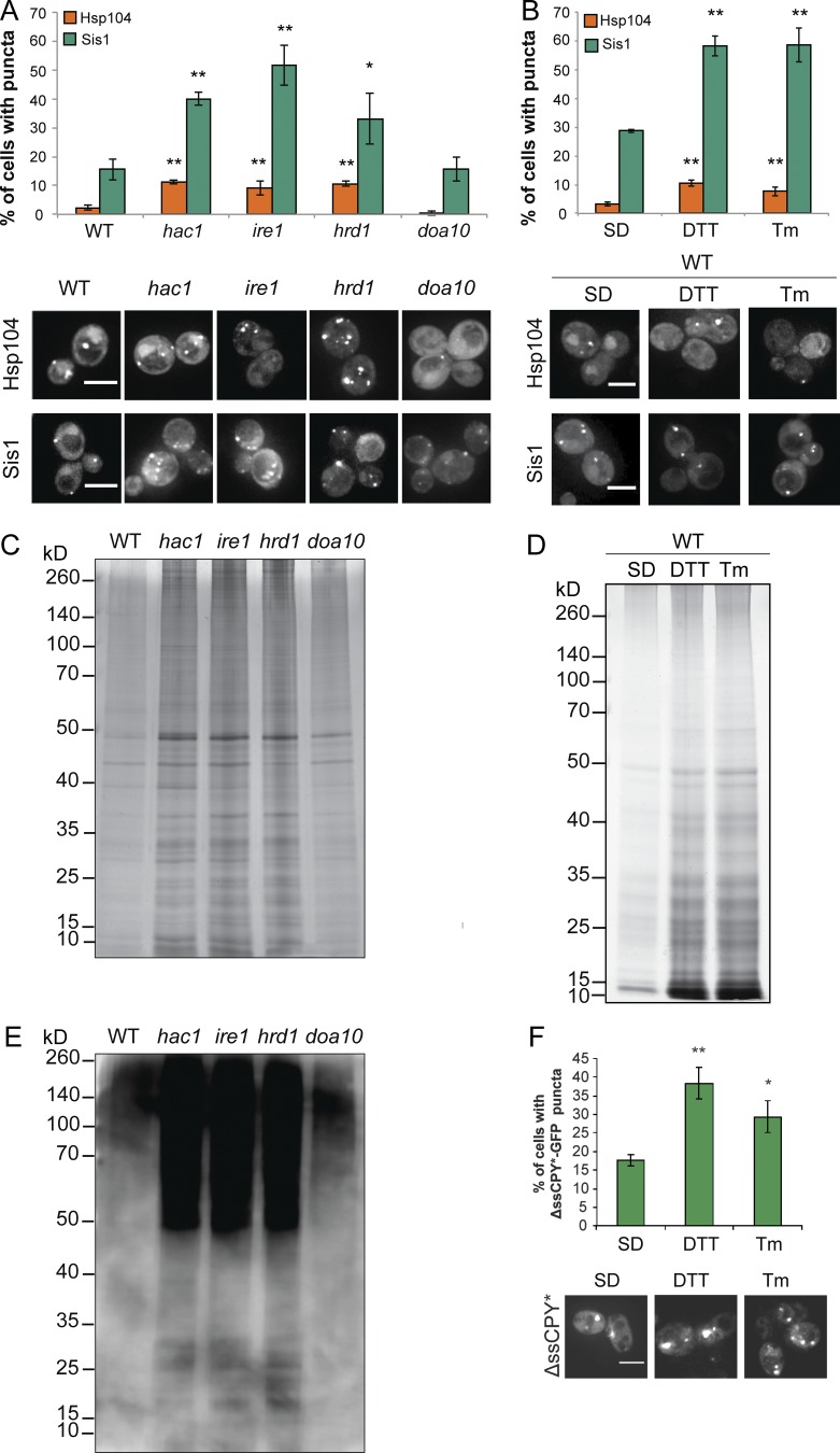
ER stress causes protein aggregation. (A) Hsp104-RFP and Sis1-GFP were visualized in wild-type, hac1, ire1, hrd1, and doa10 mutant cells and in wild-type cells exposed to DTT or Tm. Bars, 4 µm. (B) The percentage of cells containing puncta is quantified for each strain from three independent biological repeat experiments ± SD. (C and D) Silver staining of protein aggregates isolated from the same strains as in A and B. (E) Protein ubiquitination was analyzed in the isolated protein aggregates by Western blot using an α-ubiquitin antibody. (F) Examples of cells containing ΔssCPY∗-GFP puncta in a wild-type strain after DTT or Tm treatment. *, P < 0.05; **, P < 0.01; ***, P < 0.005 (n = 3).

