Figure 1.
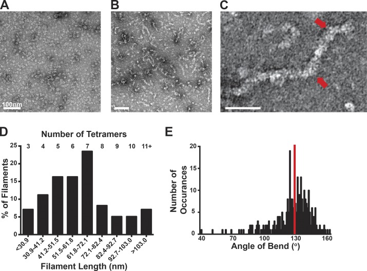
PFKL forms filaments of stacked tetramers. (A and B) TEM images of PFKL in control buffer (A) and in buffer containing 2 mM F6P (B). (C) Higher-magnification TEM image of a PFKL filament in the presence of 2 mM F6P. Red arrows highlight bends in the filament. (D) Chart of binned length determined relative to the percentage of filaments from 98 PFKL filaments measured from two separate protein preparations. The approximate number of tetramers that each bin represents is shown at the top of the chart. (E) Angle of bend determined relative to number of occurrences for a total of 290 bends measured from three separate protein preparations. The red line represents the mean of 124°.

