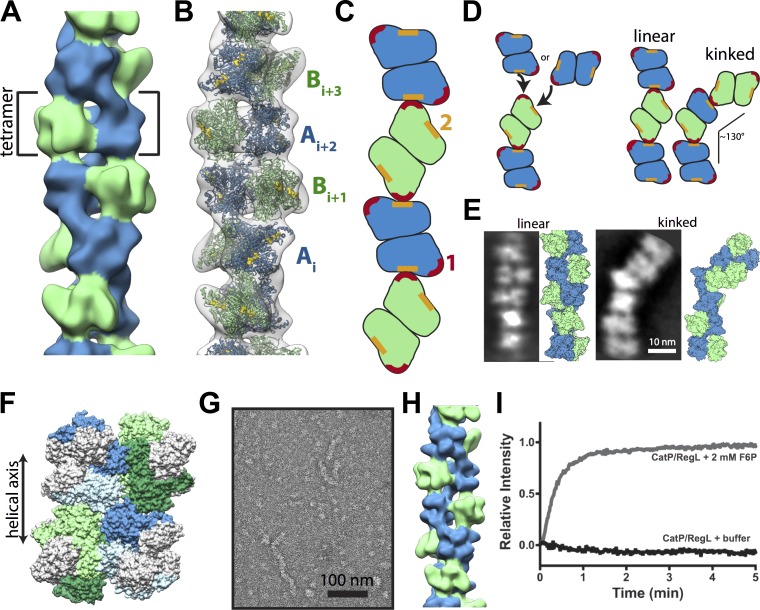
Figure 3.
Architecture of PFKL filament. (A) Negative-stain 3D reconstruction of the PFKL at 25-Å resolution. The PFKL tetramer is the repeating helical unit. Dimers on each half of the tetramer, colored blue or green, are engaged in different assembly contacts. (B) Fit of the PFKP crystal structure into the EM structure, with distinct A and B dimers labeled. (C) Diagram of longitudinal interactions between A and B dimers. (D) PFKL tetramers can add to the end of a filament through interfaces 1 or 2, resulting in either a continued linear polymer or a kink of ∼130°. (E) Examples of linear and straight filaments from reference-free means of PFKL segments with corresponding molecular models. (F) Model of CatP/RegL chimera with the catalytic domain of PFKP (gray) and the regulatory domain of PFKL (colored blue for dimer A and green for dimer B) interacting in the filament. (G) Negative-stain micrograph of the CatP/RegL in the presence of F6P. (H) Reconstruction of CatP/RegL at 25-Å resolution. (I) 90° light scattering at the 200 µg/ml CatP/RegL upon the addition of buffer alone (black line) or 2 mM F6P (gray line) at time 0. Data are representative of three determinations from two separate protein preparations.

