Figure 5.
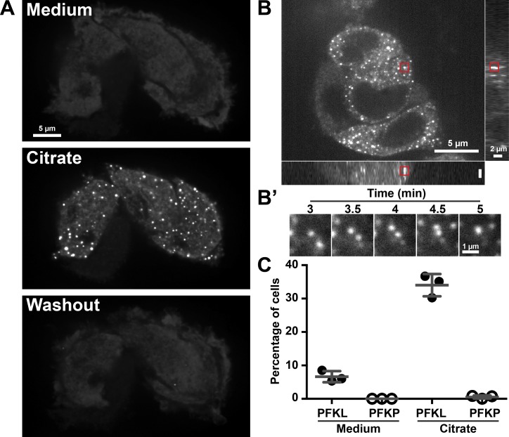
Citrate reversibly induces the formation of large PFKL-EGFP punctae. (A) Spinning-disk confocal image of PFKL-EGFP–expressing MTLn3 cells from a time-lapse sequence acquired at two frames per minute. Cells were imaged in growth medium for 5 min before the addition of 10 mM citrate. After imaging for 5 min, the citrate-containing medium was replaced with growth medium, and cells were imaged for an additional 5 min. Medium, image directly before the addition of 10 mM citrate; citrate, image 4 min after the addition of citrate; washout, image 5 min after removal of citrate-containing buffer. (B) Spinning-disk confocal z-stack of PFKL-EGFP–expressing cells 4 min after the addition of 10 mM citrate. (B′) Area of the red box in B at the time indicated after the addition of citrate. (C) Percentage of MTLn3 cells expressing either PFKL-EGFP or PFKP-EGFP showing the formation of large punctae in growth medium or 4 min after the addition of 10 mM citrate. Error bars represent means ± SD. Data are representative of three independent cell preparations.

