Figure 3.
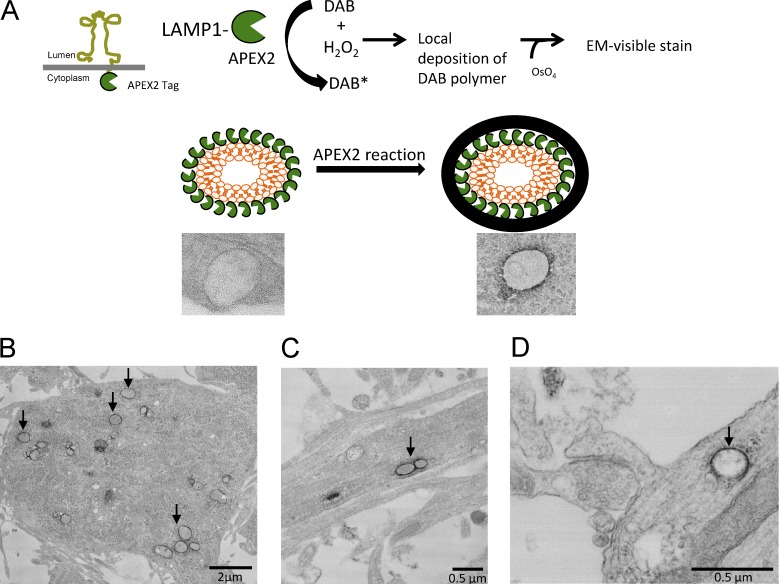
APEX2 technology for EM shows ectopic expression of LAMP1 is specific to lysosomes. (A) Schematic for how APEX2 is used to label lysosomes by EM. APEX2 was cloned on the cytoplasmic side of LAMP1 (LAMP1-APEX2) which allowed for the labeling (with minimal spread of the DAB reaction) of the outside perimeter of intact lysosomes. (B and C) Representative transmission EM images of cultured hippocampal neurons (DIV16) transfected with LAMP1-APEX2. APEX2-stained lysosomes are present in the cell body and in dendrites of neurons. (D) LAMP1-APEX2–labeled lysosome found near dendritic spines. EM image of lysosomes near a base of a dendritic spine. Arrows point to LAMP1-APEX2–positive structures.

