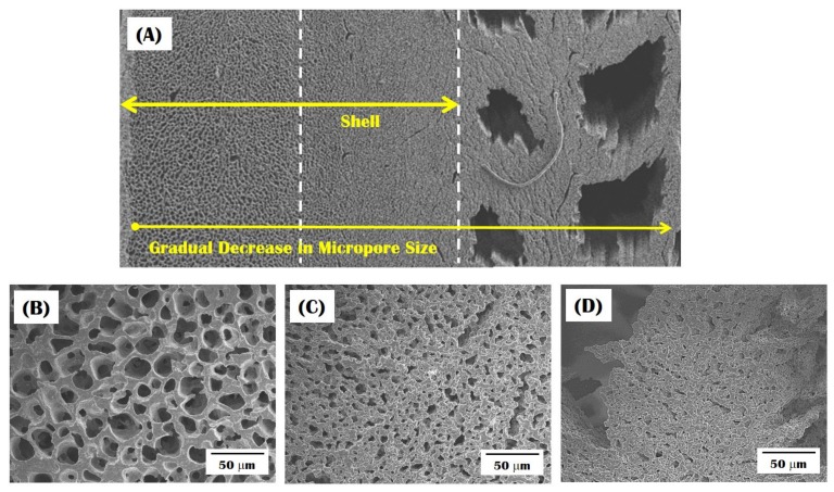
Figure 6.
Representative FE-SEM image of the bioinspired CaP scaffold showing a gradual change in microporous structure: (A) the overall structure; (B) micropores formed in the outer shell; (C) micropores formed in the region near the interface between the shell and core; and (D) micropores formed in the CaP wall in the macroporous core.

