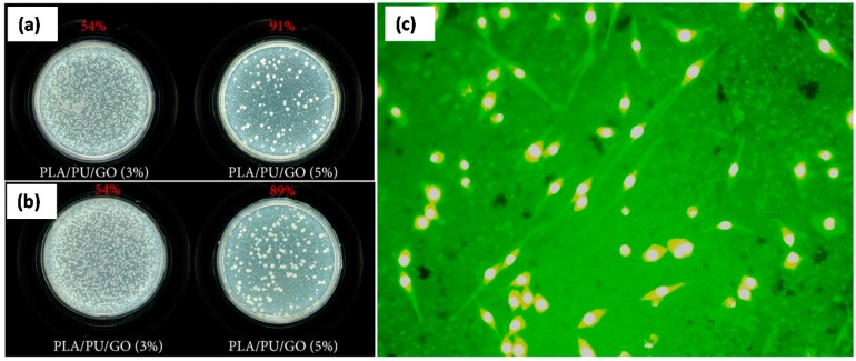
Figure 25.
Photographs of (a) S. aureus and (b) E. coli grown on PLA/PU/GO (3%) and PLA/PU/GO (5%) for 4 h, respectively; (c) Fluorescence microscopy image of MC3T3-E1 cells grown on the electrospun PLA/PU/GO (5%) nanofibers for 48 h at 37 °C. The magnification is 100× [84].

