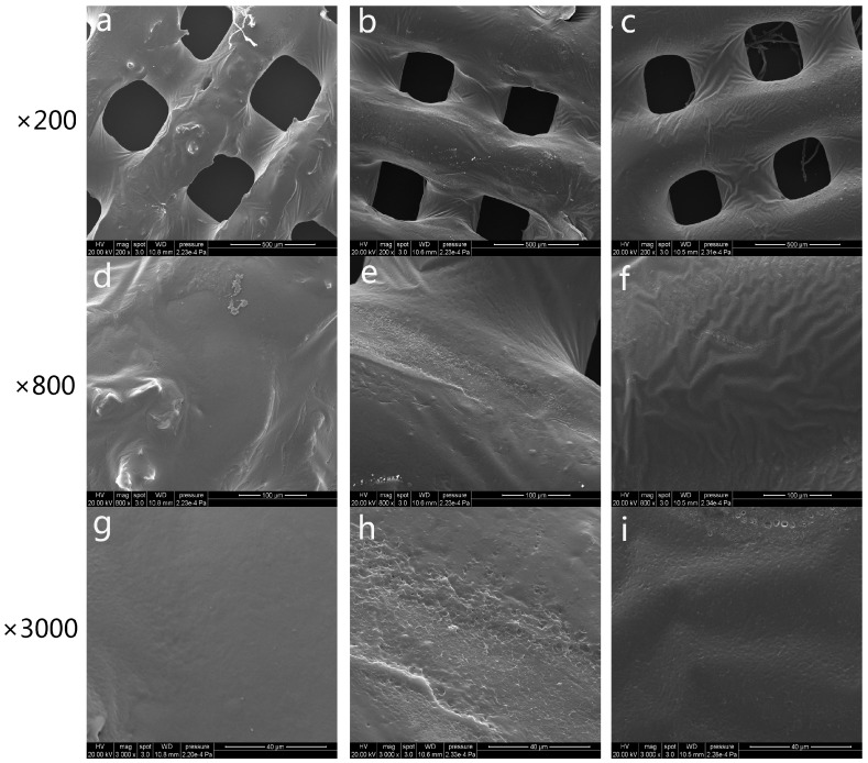
Figure 3.
Scanning electron microscope (SEM) microphotographs of freeze-dried platelet-rich plasma polycaprolactone (PRP-PCL) scaffolds (a,d,g), traditional PRP-PCL scaffolds (b,e,h), and bare PCL scaffolds (c,f,i) at ×200, ×800, and ×3000 magnification. PRP could be seen after coating with freeze-dried PRP-PCL scaffolds (a,d,g) or traditional PRP-PCL scaffolds (b,e,h). Randomly distributed PRP are visible around the surface of the scaffolds, while no PRP are visible on the bare PCL scaffolds (c,f,i).

