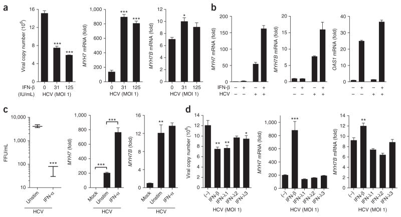
Figure 4.
Type I but not type III IFNs amplify myosin expression. Huh7 cells infected with HCV MOI 1.0 for 24 h were treated with (a) increasing doses of IFN-β, (b) 31 IU/mL IFN-β, (c) 100 ng/mL of IFN-α or (d) 100 ng/mL of IFN-λ1, IFN-λ2 or IFN-λ3 or 30 IU/mL of IFN-β. 24 h post treatment, viral copy number, MYH7, MYH7B (a–c) and OAS1 (b) expression were assessed. Reference sample for relative quantification was unstimulated HCV-infected cells. Data are from one experiment representative of two or three experiments (a–d; mean ± s.e.m., c; box and whisker plots show median values (line), 50th percentile values (box outline) and minimum/maximum values (whiskers)). Unpaired Student’s t-test was used for all statistical comparisons, *P < 0.05, **P < 0.01, ***P < 0.001. IU, international units; FFU, focus-forming units.

