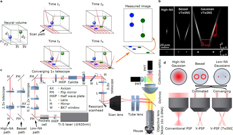
Figure 1.
vTwINS concept and design. (a) vTwINS uses a “V”-shaped PSF to image neural volumes. During scanning, the two PSF arms intersect neurons at different depths (e.g. the blue and green stylized neurons) at different time intervals. Deep neurons intersect the second arm shortly after the first. Shallow neurons take longer for the second arm to intersect. Each neuron thus appears twice, where the distance between images indicates depth. (b) Example PSFs for diffraction-limited (high-NA) TPM, and vTwINS microscopes using Bessel and low-NA Gaussian beams. (c) The vTwINS microscope consists of a beam-shaping module and a conventional two-photon microscope. The three optical paths generate the PSFs shown in (b). In the Bessel and Gaussian (low-NA) vTwINS paths, lenses adjust the PSF’s axial extent, and a birefringent block (calcite) splits the beam in two and sets the PSF angle. (d) The back aperture illumination profiles for the three paths in (c). In the high-NA (conventional TPM) path, the overfilled back aperture is focused to a point. In the Bessel and low-NA Gaussian paths, two beams are focused to form each arm of the PSF. The beam divergence is adjusted with the 1× telescope before the calcite block to separate the two arms of the X-PSF and form the V-PSF.

