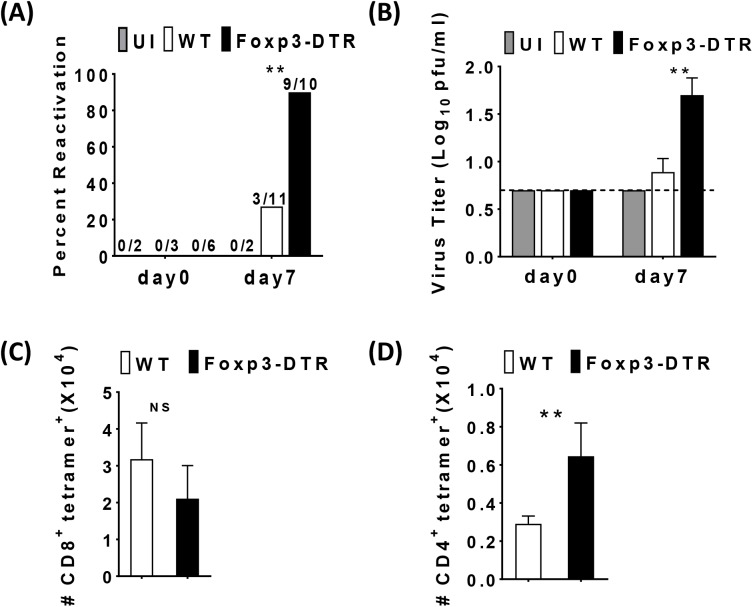
Fig 4. Treg are required to prevent MCMV reactivation and MCMV-specific CD4+ T cells in the SG.
5–6 week old WT C57BL/6 and Foxp3DTR mice were inoculated with1× 106 pfu of MCMV. 8 months post-MCMV infection, both groups were injected with Diphtheria toxin (DT) on day 0, 3, 6 and sacrificed on day 7. Bar graph (A) shows the percentage of WT C57BL/6 uninfected control (UI, N = 2 day 0, N = 2 day 7, gray bar), MCMV infected (N = 3 day 0, N = 11 day7 white bars) and Foxp3DTR (N = 6 day 0, N = 10 day 7, black bars) mice positive for virus replication in the SGs before Treg depletion (indicated as day0) and day7 post Treg depletion with the numbers of positive mice in each group shown above the bars. (B) Bar graph shows the average viral titer of individual SGs of MCMV infected mice before Treg depletion (indicated as day0) and day7 post Treg depletion as described above (mean+SEM). The presence of replicating virus was detected by plaque assay. Samples with no detectable virus were assigned a titer of 0.7 log pfu/ml, the limit of detection for the plaque assay as indicated by the dashed line. Data is representative of five independent experiments. Statistical analysis, *p ≤ 0.05, **p ≤ 0.01 (Student’s t tests) comparing infected mice. Single cell suspensions were generated from the SGs of MCMV infected mice and were stained first with MCMV-tetramers, M38 (CD8), M25 (CD4) and then cells were surface stained for CD8, CD4, and analyzed with flow cytometry. Bar graph (C) shows the total number of M38-specific CD8 T cells in the SGs (mean+SEM). WT C57BL/6 (N = 4). Foxp3DTR (N = 5). Bar graph (D) shows the total number of M25-specific CD4 T cells in the SGs (mean+SEM). WT C57BL/6 (N = 6). Foxp3DTR (N = 8). Statistical analysis, *p ≤ 0.05, **p ≤ 0.01 (Student’s t test).

