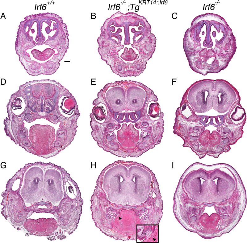
Figure 3.

Rescue embryos have oral adhesions and cleft palate at birth. Head coronal sections of perinatal pups stained with H&E. A–C) anterior, D–F) middle and G–I) posterior oral cavities of wildtype (A, D, G), rescue (B, E, H) and knockout (C, F, I) pups. The oral cavity of rescue pups is less severely affected overall but oral adhesions persist bilaterally in the anterior oral cavity (B) and in the middle oral cavity between the maxilla and mandible at the tooth germ (E). In the posterior palate of rescue embryos (H), adhesions and a fusion (black arrow head, enlarged with inset) is observed between the palate and the tongue. (A) Scale bar = 500 μm. T, Tongue; PS, palatal shelves.
