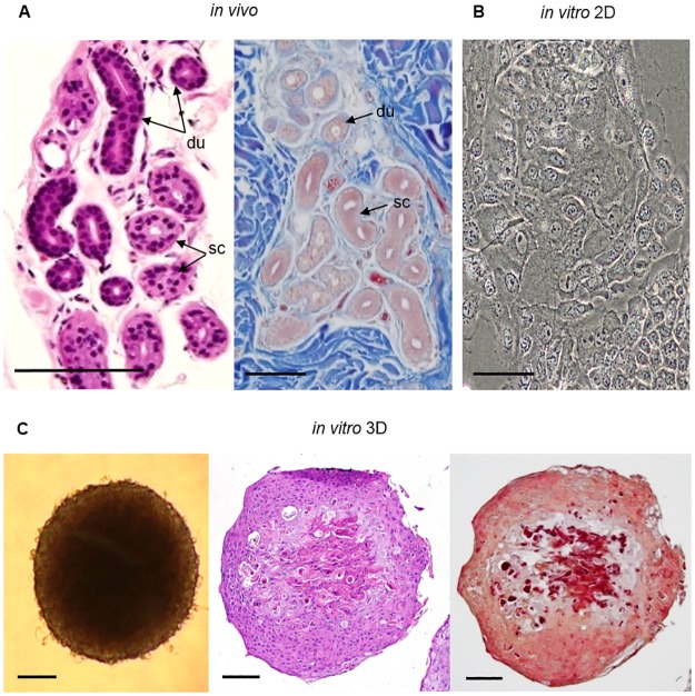
Fig 1. Histological analysis of human eccrine sweat glands, monolayer cells and 3D cell culture models in vitro.
(A) H&E and azan staining of paraffin axillary skin tissue sections visualizing the morphology of native eccrine SG’s with secretory coil portions (sc) and ductal portions (du) of the gland. (B) Bright-field microscopy of primary SG cells in 2D culture with their typical ‘cobblestone’ morphology is shown. (C) Bright-field microscopy and H&E-/azan-stained sections of a differentiated SG 3D model with irregularly arranged, multilayered cells and less dense cells in the center of the spheroid. Collagen in blue could not be detected in azan staining, but acidic structures like nuclei or matrix were stained in red or dark red. Scale bars 100 μm.

