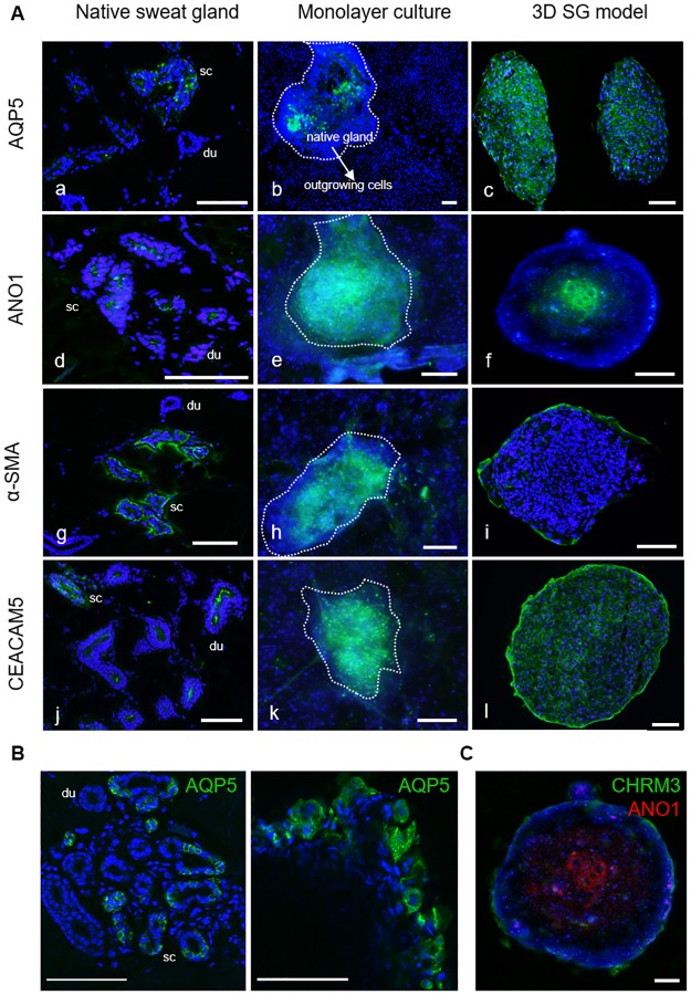
Fig 4. In vitro characterization of key marker proteins AQP5, ANO1, α-SMA, and CEACAM5 in cultures of the SG by immunofluorescence staining.
(A) Stained secretory coil (sc) and duct (du) sections of native human sweat glands were analyzed in axillary skin tissue (a,d,g,j) compared to in situ glands (dashed lines) with outgrowing cells in 2D cultures (b,e,h,k) and 3D models (c,f,i,l). Polarization and differentiation of spheroids could be shown with luminal ANO1 (f) and basolateral α-SMA staining (i). Outgrown SG cells showed decreased marker expressions in the 2D cultures. Nuclei stained with DAPI. (B) By confocal imaging, the localization of AQP5 was investigated in sections of native skin tissue with eccrine sweat glands and in whole mount-stained 3D models. In vivo and in vitro, AQP5 could be shown in the cytoplasm, in the basolateral and the luminal membrane of secretory coil cells and in cells of the 3D model. (C) Localization of ANO1 and CHRM3 was analyzed to investigate the apical-basal polarity in the 3D model. Here, the whole mount-stained 3D model indicated this histoarchitecture with luminal ANO1-staining in the center and basolateral CHRM3-staining in the outer layer of the sphere. Scale bars 100 μm.

