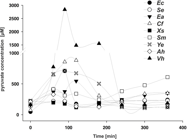
Fig 6. Extracellular concentrations of pyruvate during growth of the indicated γ-proteobacterial species in LB medium are plotted against time after inoculation.
At the times indicated, cells were harvested, and pyruvate levels in the cell-free supernatant were quantified by hydrophilic interaction liquid chromatography. All experiments were performed in triplicate, and the error bars indicate the standard deviations of the means. Abbreviations as in Fig 4.

