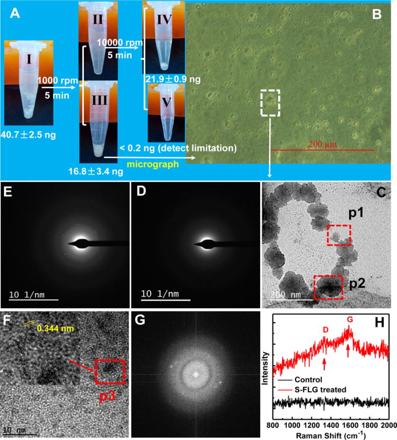
Figure 6.

(A) S-FLG content in intestinal epithelial cells collected from the zebrafish of the S-FLG treated group, background radioactivity was subtracted. Tube I, gut tissues (after removal of the intestinal contents) were incubated with enzyme solution (collagenase and dispase). Tube II, mixture after the removing of cellular precipitate. Tube III, cellular precipitate. Tube IV, residual tissues. Tube V, mixture solution. Data are presented as mean ± standard deviation (n=5).; (B) microscope of the obtained cells; (C) TEM image of the intestinal epithelial cells; (D) and (E) SADPs taken from p1 (control) and p2 positions, respectively. Note: six fold symmetry was observed in (E); (F) High resolution TEM image taken from p2 position in (C) (the red arrow pointed to an enlarged view of p3 position); (G) Fourier transfer spectra of Figure F; (H) Raman spectra of the sectioned intestinal epithelial cells of zebrafish treated by S-FLG.
