
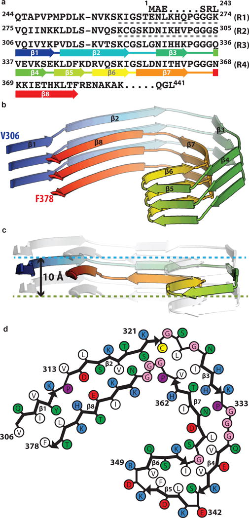
Figure 3. The common protofilament core.
a. Sequence alignment of the four microtubule-binding repeats (R1–R4) with the observed eight β-strand regions coloured from blue to red. The sixteen residues from R1 or R2 that may form an additional, less-ordered β-sheet are indicated with grey dashed lines. b. Rendered view of the secondary structure elements in three successive rungs. c. As in b, but in a view perpendicular to the helical axis, revealing the differences in height along the helical axis within a single molecule. d. Schematic view of the protofilament core.