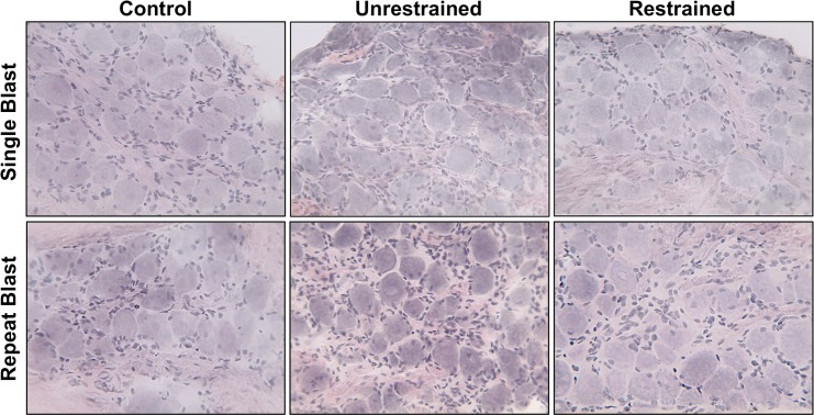
Fig 2. H&E staining of trigeminal ganglion (TG) sections.
Frozen sections of TGs, from all animal treatment groups, were subjected to H&E staining to assess ganglion structure and anatomy. Staining revealed large sensory cell bodies surrounded by satellite cells. Results are representative images of all animal groups (40X magnification).

