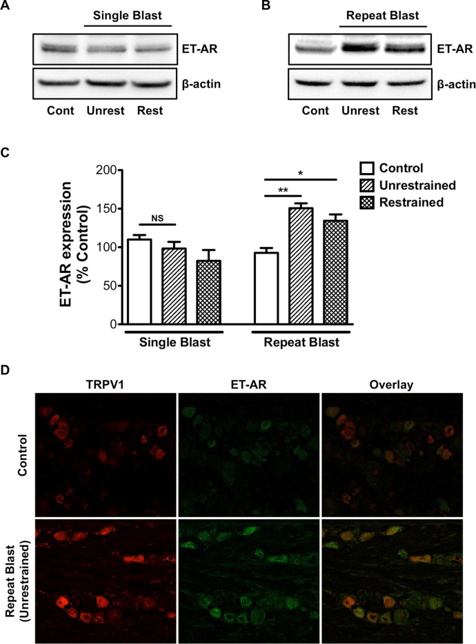
Fig 4. Repeat blast exposure results in increased TRPV1 and ET-A co- expression.
Western blot and immunofluorescence analysis was performed on TGs harvested from animals that were exposed to either a single or repeat low-level primary or tertiary low-level blast. (A, B) TGs were homogenized and whole cell lysates (50 μg) were assessed for ET-A protein expression. (C) Quantification of ET-A expression in unrestrained and restrained animals following exposure to a single or repeat blast. (D) Immunofluorescence staining was completed on frozen sections of TGs subjected to repeat, tertiary blast. Tissues were subjected to staining with anti-TRPV1 (1:250) and anti-ET-A (1:250) antibodies and probed with Alexa Fluor 568 and 488 secondary antibodies, respectively. Representative images of an n = 4 for both control and repeat blast animals. NS, not significant, *p<0.05 and **p<0.01, one-way ANOVA with Tukey post-hoc test.

