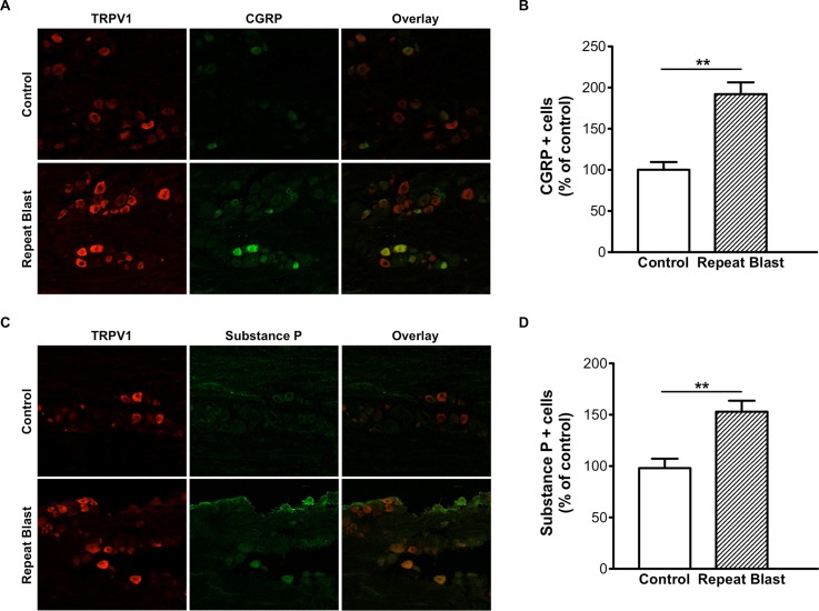
Fig 7. Increased CGRP and SP expression in TG sensory neurons exposed to repeat, tertiary blast.
Immunofluorescence analysis was performed on frozen TG sections harvested from control and repeat, tertiary (unrestrained) blast exposed animals. Tissues were subjected to staining with anti-TRPV1 (1:250), anti-CGRP (A) (1:250) and anti-SP (C) (1:250) antibodies and probed with the appropriate Alexa Fluor 568 and 488 secondary antibodies. (B, D) Quantification of CGRP- and SP-positive cells as percent of control animals, respectively. Paired t-test, **p<0.01.

