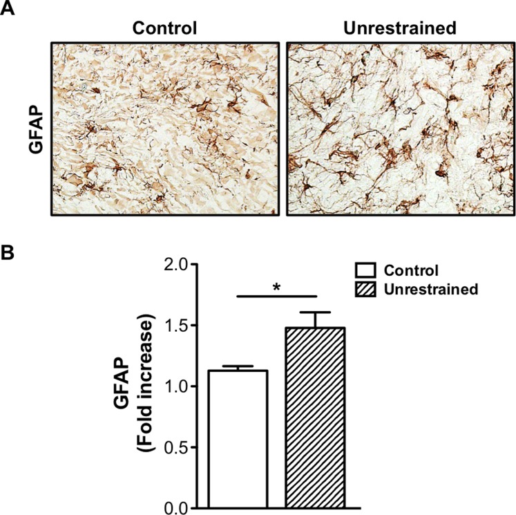
Fig 8. Repeat low-level blast increases GFAP expression in the trigeminal ganglion.
(A) Immunohistochemical staining was performed on frozen TG sections harvested from control animals and unrestrained animals exposed to repeat, tertiary blast. Tissues were subjected to staining with anti-GFAP (1:1000) and then probed with the appropriate secondary biotinylated antibody. (B) Quantification of GFAP expression in TG homogenates from control and repeat, tertiary blast treatment groups. Data represented as fold increase in GFAP expression as compared to control. NS, not significant, *p<0.05, one-way ANOVA with Tukey post-hoc test.

