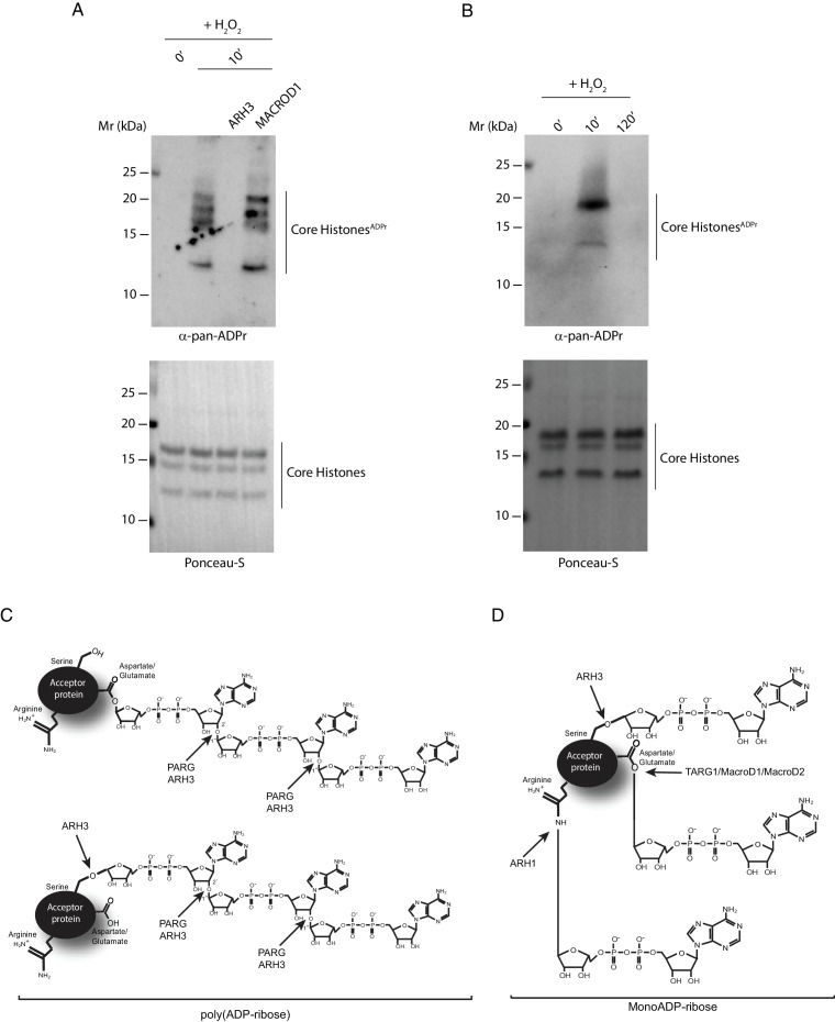
Figure 6. ARH3 as a tool for recognizing histone Ser-ADPr.
(A) Core histones were purified from U2OS cells treated with H2O2 for the indicated time points. Recombinant ARH3 or MACROD1 or reaction buffer was added to histones purified from 10 min H2O2-treated cells. After treatment, proteins were separated by SDS-PAGE, analysed by western blot and probed for pan-ADPr. Ponceau-S staining was used as loading control. (B) Purified core histones from U2OS cells treated with H2O2 for the indicated time points were separated by SDS-PAGE, analysed by Western blot and probed for pan-ADPr. Ponceau-S staining was used as loading control. (C and D) A schematic representation of the specificity of the ADP-ribosylhydrolases PARG, ARH3, TARG1, MACROD1, MACROD2 and ARH1 for PARylated (C) and MARylated (D) proteins.

