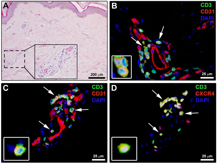
Fig 7. Angiogenic T cells (Tang) are present in systemic sclerosis (SSc) dermal perivascular inflammatory infiltrates.
(A) Representative microphotograph of skin sections from SSc patients stained with hematoxylin and eosin. Perivascular inflammatory cells from the boxed area are shown at higher magnification in the inset. (B) Representative fluorescence microphotograph of skin sections from SSc patients double immunostained for the pan-T lymphocyte marker CD3 (green) and CD31 (red), and counterstained with 4′,6-diamidino-2-phenylindole (DAPI; blue) for nuclei. Arrows indicate CD3+CD31+ T lymphocytes. (C, D) Representative fluorescence microphotographs of serial skin sections from SSc patients double immunostained for CD3 (green) and CD31 (red; C) or CXCR4 (red; D). Arrows indicate CD3+CD31+CXCR4+ T lymphocytes (Tang). Insets: higher magnification views of immunopositive lymphocytes from the respective panels. Scale bars are indicated in each panel.

