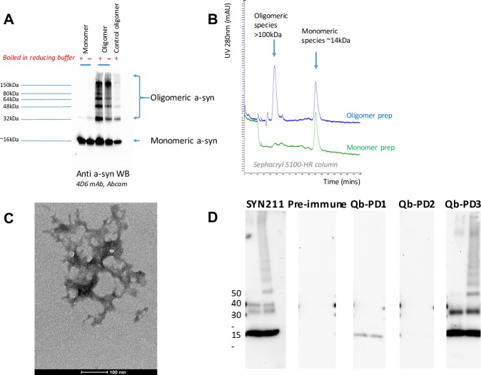
Fig 4. Characterisation of a-syn oligomer preparations and recognition of these oligomers by IgGs from Qb-PD vaccinated mice.
(A) Western blot (WB) of monomer and oligomer a-syn preparations reduced with (+) or without heating (-) probed with 4D6 a-syn antibody (1:2000). (B) A 100 μL sample of each preparation was passed through a Sephacryl S100-HR column, displaying over-layered chromatograms to compare UV (280nm) elution profiles. (C) Negative stained TEM of tangled oligomer preparation (scale bar, 100nm). (D) WB on 0.1 μg of full-length recombinant a-syn monomers (left lane) and a-syn oligomers prepared with 4-hydroxy-2-nonenal (HNE) (right lane). WB were probed using SYN211 a-syn antibody (1:5000) and IgGs (1:500) from vaccinated male and female SNCA-OVX mice. These vaccinated mice received 20 μg of Qb, Qb-PD1 or Qb-PD3 every two weeks for a month, followed by a monthly injection (total duration of immunisation: 2 months). Pre-immune refers to sera of mice collected prior to first immunisation at d0.

