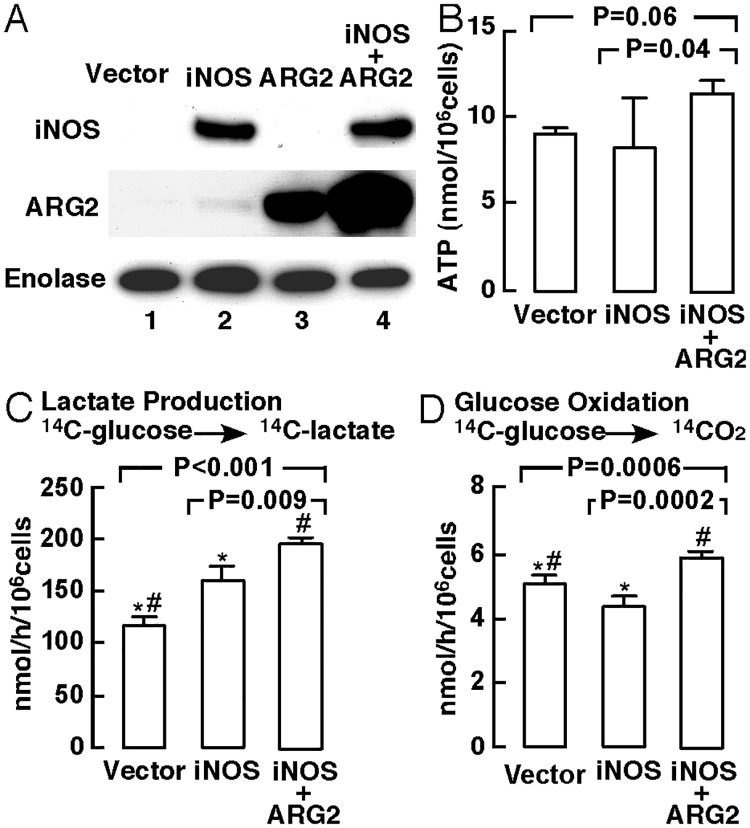
Fig 2. Protein expression and bioenergetics of bronchial epithelial cells with iNOS expression.
(A) Western analyses of BET1A cells transfected with iNOS vector, ARG2 vector, co-transfected with iNOS+ARG2, or control vector (n ≥ 3 replicate experiments). Enolase was used as a loading control. (B) ATP production determined in BET1A cells transfected with iNOS vector, co-transfected with iNOS+ARG2, or control vector (n ≥ 3 replicate experiments). (C–D) Radioisotope studies of glucose metabolism in BET1A cells with iNOS expression. The rates of production of lactate (C) and oxidation of glucose (D) using radioactive glucose tracer were analyzed in BET1A cells transfected with iNOS vector, control vector, or co-transfected with iNOS+ARG2 vectors (n ≥ 3 replicate experiments). *P < 0.05, iNOS-expressing cells vs. control vector-transfected cells; #P < 0.05, iNOS+ARG2 co-transfected cells vs. control-transfected cells.

