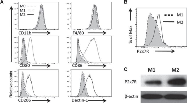
Figure 4. Analysis of cell surface markers expressed by polarized M1 and M2 cells.

(A) Mouse bone marrow–derived macrophages were polarized into M1 cells (cultured in LPS and IFN-γ) or M2 cells (cultured in IL4- and IL-13), and macrophages cultured without polarizing cytokines were referred as M0 cells. The polarized macrophages were analyzed for expression of CD11b, F4/80, CD80, CD86, CD206, and Dectin-1 expression by FACS. (B) The FACS plot showing the expression of P2x7r by polarized M1 and M2 cells. (C) Western blot showing P2x7r protein expression in polarized M1 and M2 cells. Representative data of one of three experiments are shown. FACS, fluorescence-activated cell sorting; P2x7r, purinergic receptor P2X7.
