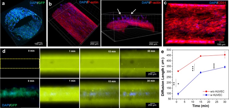
FIG. 3.
Confocal microscopy images of (a) the integrity of the GFP-HUVEC layer in the construct with nuclei staining after 2 days and (b) top and cross-sectional views of F-actin/nuclei staining after 7 days which showed endothelial cell invasion and sprouting into the surrounding GelMA hydrogel (arrows). (c) Top view of a HUVEC layer immunostained for CD31 (red) and 4′,6-diamidino-2-phenylindole″ (DAPI) (blue) after 2 days in culture. (d) Diffusion profiles of Rhodamine 6G into the microchannel endothelialized without (upper row) and with (lower row) GFP-HUVECs at several time points. The channel's diameter was 500 μm. (e) Diffusion length of Rhodamine 6 G with and without the endothelial cell barrier. Fluorescence intensity was measured at the center of the channel. The barrier delayed the diffusion of the dye (*** p < 0.001).

