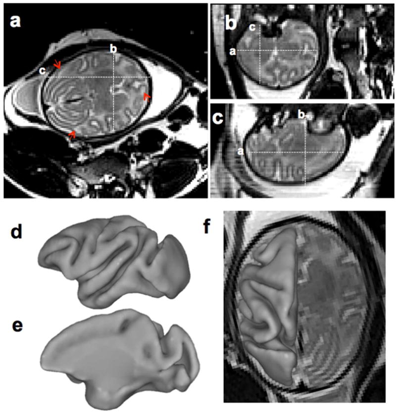
Figure 3. In utero T2-weighted control fetal brain images and cortical surface at 139dGA.

T2-weighted image was acquired using HASTE along the axial direction. From axial (a), coronal (b), and sagittal (c) view, tissue contrast between cortical grey matter, fetal white matter and deep nuclei is evident (a–c). Heterogeneous signal intensity within the developing cerebral cortex is present at this developmental stage (A, arrowheads). The cerebral cortical surface was generated from segmentations of the T2-weighted image (d–f). At this developmental stage, all primary and secondary sulci and gyri present in adult cortex are identifiable.
