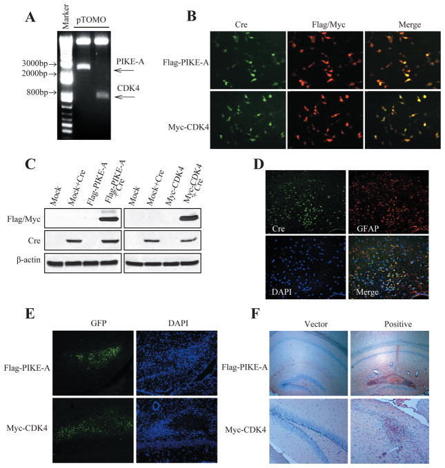
Figure 1. Cre recombinase-dependent expression of Flag-PIKE-A/Myc-CDK4 by pTOMO PIKE-A/CDK4 lentiviral vector (LV) in vitro and in vivo.
A. Plasmids confirmation of pTOMO-Flag-PIKE-A and pTOMO-Myc-CDK4. pTOMO constructions were digested by BamHI an EcoRV. B. Immunocytochemistry showing Flag-PIKE-A/Myc-CDK4 expression induced by Cre recombinase. HEK293 cells were infected with pTOMO-Flag-PIKE-A/pTOMO-Myc-CDK4 LVs with Cre-expressing LVs. After fixation, the cells were stained with the indicated antibodies. C. Western blot showed Flag-PIKE-A/Myc-CDK4 expression induced by Cre recombinase. HEK293 cells were infected with pTOMO mock LVs or pTOMO PIKE-A/CDK4 LVs with or without Cre-expressing LVs, and the cell lysates were processed for western blotting with indicated antibodies. β-actin detection was used as a loading control. D. Confocal images of Cre recombinase specifically expressed in GFAP+ cells in GFAP-CreER mice. Brain sections were analyzed by immunofluorescence staining, Cre and GFAP expressed cells were stained as described in Experimental procedures. E. Tomo PIKE-A/CDK4 LVs were successfully injected into hippocampus (HP). Images were taken 10 days after injection of pTOMO PIKE-A/CDK4 LVs into HP. GFP signals (green) indicated the infected cells. F. Immunohistochemistry images showing Tomo PIKE-A/CDK4 were successfully expressed in mouse brains. Staining was taken 10 days after injection of pTOMO PIKE-A/CDK4 LVs into HPs. The brown cells showed the positive cells.

