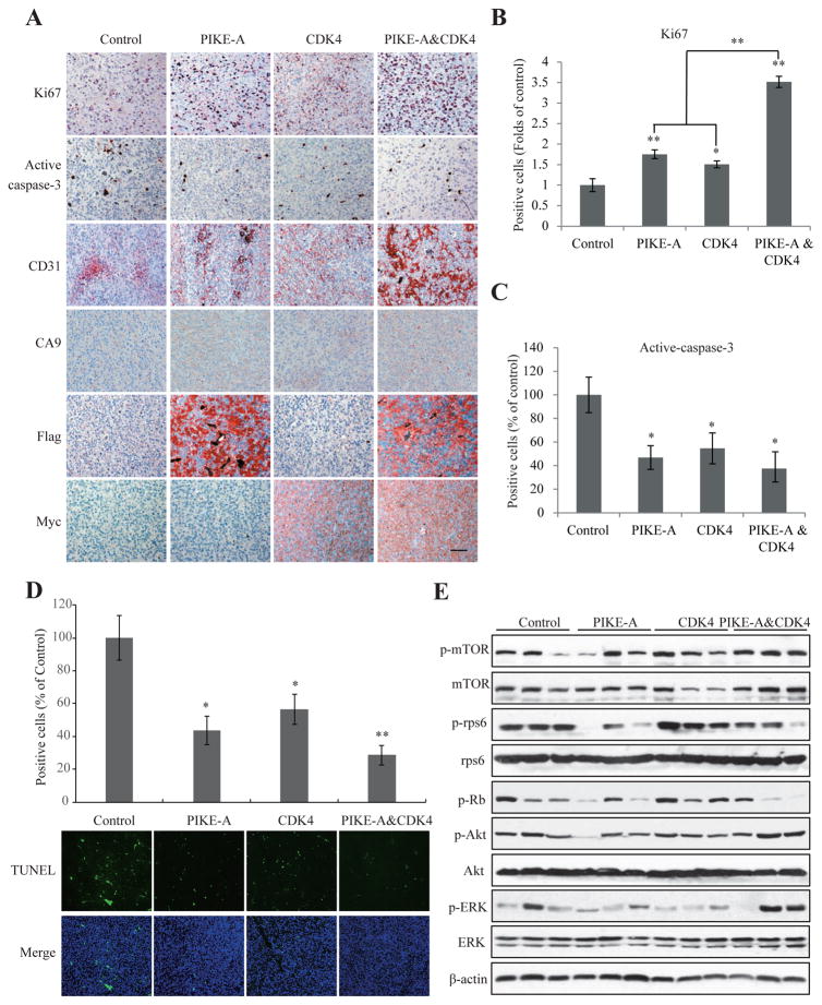
Figure 3. PIKE-A and CDK4 additively promote tumor growth in vivo.
A. Immunohistochemistry analysis of cell proliferation, apoptosis and angiogenesis using Ki67, active-caspase-3, CD31, and CA9 in the brain tumors from indicated groups. Scar bar, 20 μm. B & C. Quantification of Ki67 (B) and active-caspase-3 (C) positive cells in the slides of brain tumors in different groups. D. Apoptosis detection/quantification using by TUNEL staining. Three fields were averaged in each tumor, and the averages for each animal yielded the final mean ± SEM (*P < 0.05, **P < 0.01, two-tailed Student’s t test, n = 3). E. Signaling characterization in the tumors formed in the brains infected by indicated lentivirus. Brain tumors were collected, lysed and equal amount of proteins in each sample were analyzed by western blotting with the indicated antibodies.

