
Figure 1. HBEGF promotes anchorage-independent growth of immortal astrocytes.
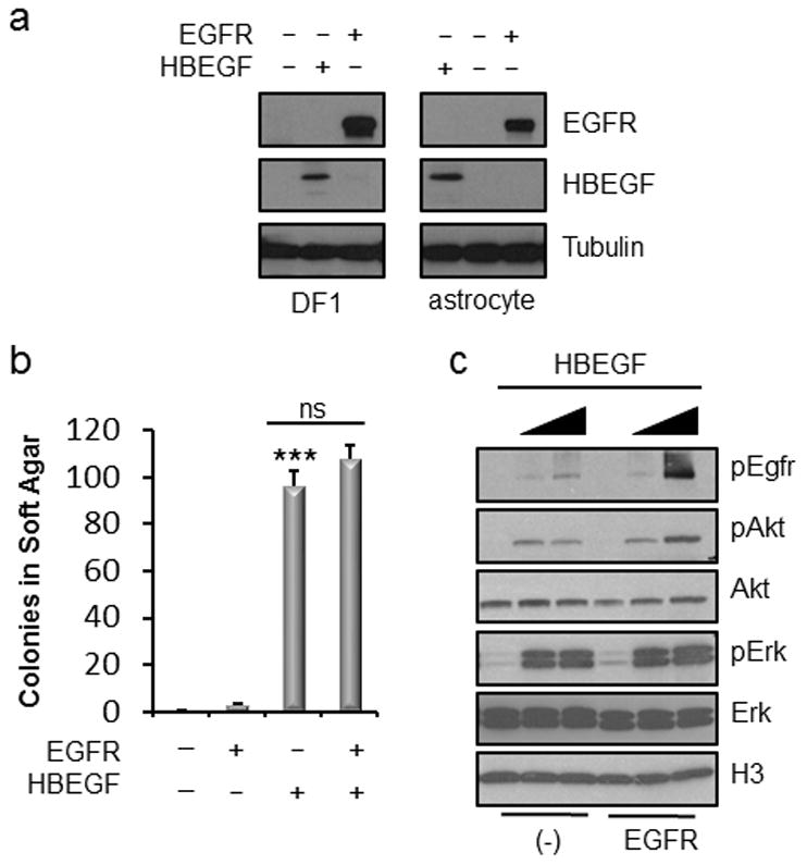
(a) Expression of EGFR and HBEGF in DF-1 cells and Nestin-TVAInk4a/Arf lox/lox mouse astrocytes. (b) Soft agar colony formation of Ink4a/Arf lox/lox astrocytes expressing exogenous EGFR and/or HBEGF compared with RCAS-Cre infected control cells. Data is represented as mean ± S.E.M. Using a two-tailed Student’s t test, the number of colonies formed were compared to control and statistical significance is shown by the following key: *** P < 0.0005 with not significant (ns) denoting P > 0.05. (c) Western blot analysis of downstream effector pathways in Ink4a/Arf lox/lox astrocytes expressing either endogenous (−) or exogenous (EGFR). HBEGF was added to serum-free media at two different concentrations (0.5 ng/ml and 5.0 ng/ml).