
Figure 4. Both Egfr and Axl mediate HBEGF signaling.
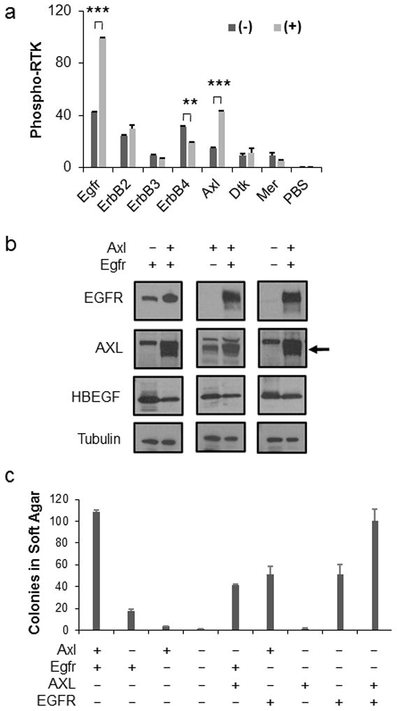
(a) Phosphorylation of multiple RTKs was simultaneously assayed in isogenic astrocytes in the presence (+) or absence (−) of HBEGF. A subset of RTKs analyzed representing the ErbB and TAM family members are shown. Densitometry of the array was used to determine the amount of RTK phosphorylation. Data are represented as mean ± S.E.M. Statistical significance is shown by the following key: ** P < 0.005, *** P < 0.0005. (b) Endogenous Axl and/or Egfr expression was eliminated using CRISPR-mediated gene editing (−) in Nestin-TVA;Ink4a/Arf lox/lox astrocytes. Human AXL and EGFR were expressed following RCAS-mediated infection for rescue (+). The arrow represents the AXL band. Blots were probed with tubulin antibody as a loading control. (c) Astrocytes from (b) were assayed for soft-agar colony formation to determine the functional significance of Axl and Egfr loss. Data are represented as mean±S.E.M. Cells lacking either Axl, Egfr, or both had significantly fewer colonies than the unmodified parental cells (P < 0.0005). Rescue with human AXL or EGFR in the corresponding single knockout cells was significant (P < 0.005). There was no significant difference between EGFR rescue in the single versus double knockout cells; however, a significant difference was observed between AXL rescue in the single versus double knockout cells (P < 0.0005). Expression of both AXL and EGFR completely rescued the double knockout cells as no significant difference was observed between the double rescue and the control parental cells.