
Figure 7. Suppression of HBEGF expression prolongs survival in vivo.
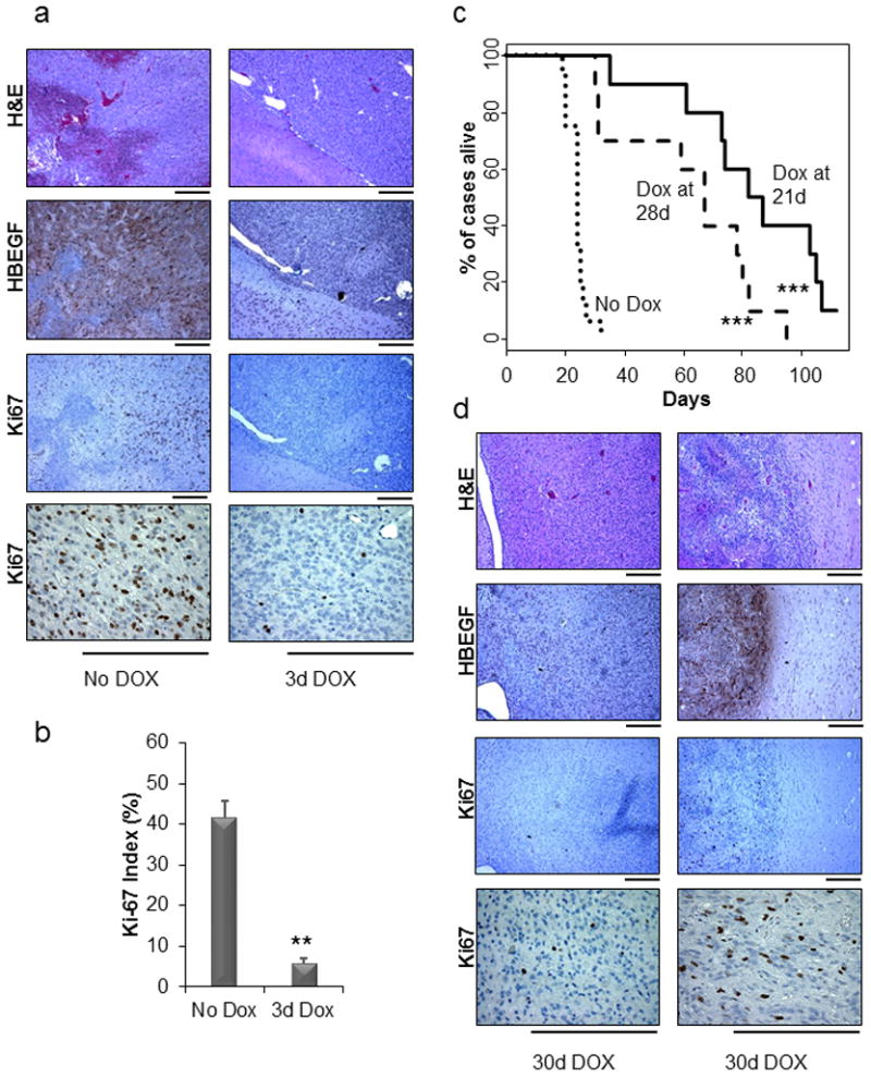
(a) Immunohistochemical analysis of HBEGF and Ki67 in brain tissue isolated from Nestin-TVA;Ink4a/Arf lox/lox;Ptenlox/lox mice injected with RCAN-TRE-HBEGF, RCAS-tet-off, and RCAS-Cre that were either untreated or treated with doxycycline for 3 days after being weaned at 21 days of age. The two samples are from the same timepoint of 3 days. Scale bar represents 300 μm. (b) The Ki67 staining was quantified using two representative tumors for each cohort. For each tumor, the average of four high-power fields is shown. Data are represented as mean ± S.E.M (** P < 0.005). (c) Kaplan-Meier survival analysis of Nestin-TVA;Ink4a/Arf lox/lox;Ptenlox/lox injected with RCAN-TRE-HBEGF, RCAS-tet-off, and RCAS-Cre that were not treated (n = 16, round dash), treated starting at 21 days of age (n = 10, solid line), or treated starting at 28 days of age (n = 10, short dash). The Dox treated cohorts were compared with the untreated cohort (*** P < 0.0005). (d) Immunohistochemical analysis of regressed tumors using antibodies against HBEGF and Ki67 with corresponding H&E. Scale bar represents 300 μm. Both regressed tumors are from two different mice that were treated for 30 days with doxycycline.