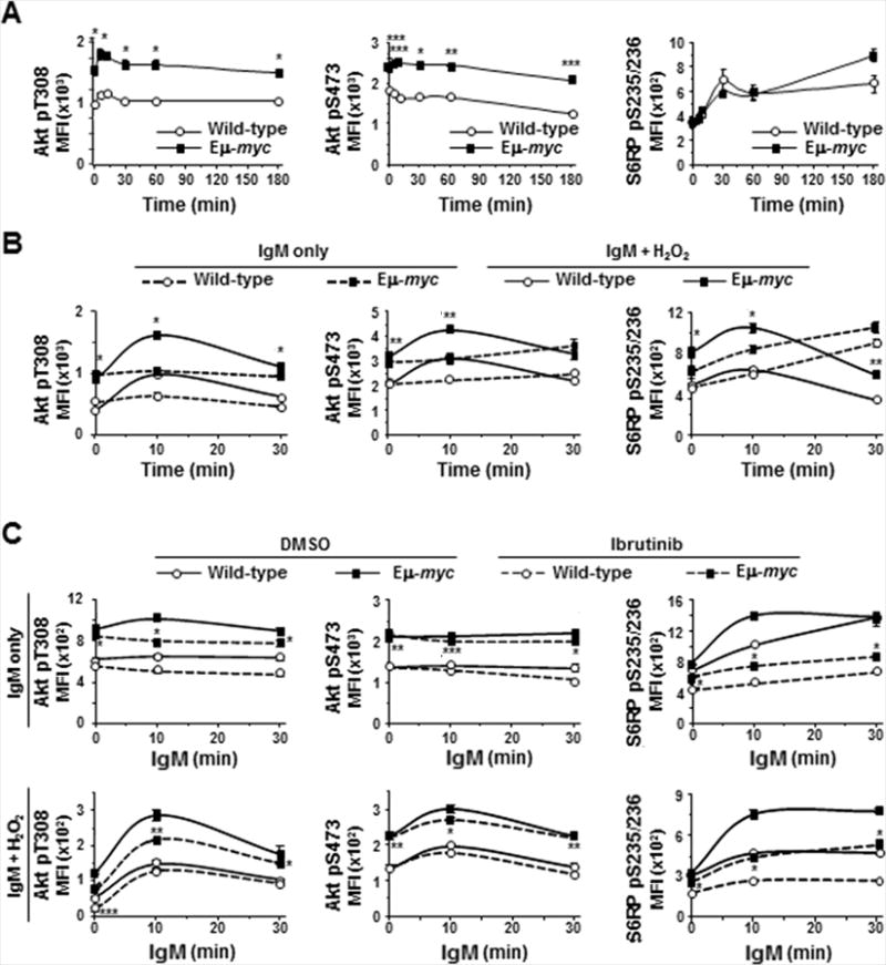
Figure 6. Signaling through PI3k/Akt is enhanced in Eμ-myc B cells and inhibited by ibrutinib.

Phosphorylation of Akt and S6 ribosomal protein (S6RP) in splenic B cells was measured by intracellular phospho-flow cytometry at intervals after IgM ligation without (A, upper panels C) or with (B, lower panels C) addition of H2O2 in the absence (A, B) or presence (C) of ibrutinib. Each protein was measured in at least three experiments with 2–3 mice of each genotype. Mean fluorescence intensities (MFI) from one representative experiment are shown; p-values compare phospho-protein levels in Eμ-myc and wild-type B cells in IgM-stimulated (A), IgM-stimulated + H2O2 (B, solid lines), ibrutinib-treated (C, dashed lines); *p≤0.01, **p≤0.03, and ***p≤0.05 (p values for other comparisons not denoted).
