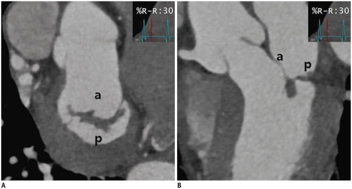
Fig. 8. Cardiac CT images after MV repair due to mitral regurgitation caused by MV prolapse.
En face view (A) and three-chamber view (B) at level of mitral valve during systolic phase showing well functioning mitral valve without coaptation defect. a = anterior leaflet of MV, p = posterior leaflet of MV

