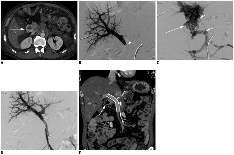
Fig. 1. 53-year-old man with hematochezia.
Patient underwent pylorus-preserving pancreaticoduodenectomy due to pancreatic cancer 737 days ago.
A. Axial image shows varix in afferent jejunal loop (arrow). B, C. Direct portogram (B) via transhepatic approach shows occlusion of main portal vein (arrowhead). Superior mesenteric venogram (C) demonstrates extensive collateral channels along afferent jejunal loop (arrow) and segmental occlusion of portal vein (smaller arrows). D. Direct venogram after deployment of stent (12 mm in diameter and 80 mm in length) shows disappearance of collateral channels and opacification of both portal veins. E. Follow-up CT performed 60 days after portal stenting shows patent stent and disappearance of varix in afferent jejunal loop (arrow).

