. 2017 Jul 31;58(5):899–909. doi: 10.3349/ymj.2017.58.5.899

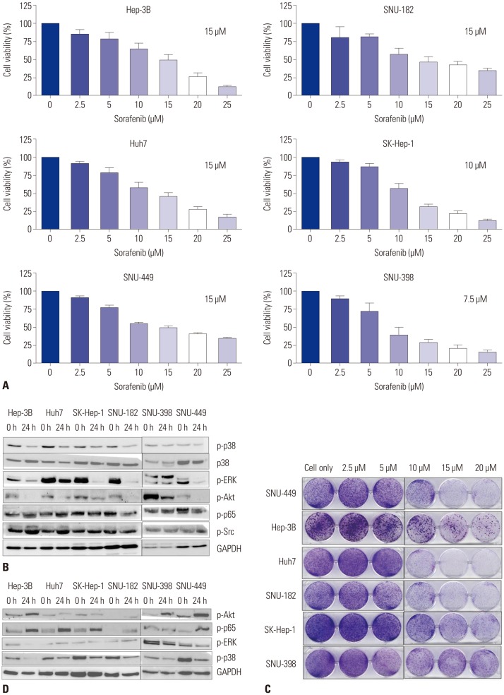
Fig. 1. Effect of sorafenib on different HCC cell lines. (A) Hep-3B, Huh7, SK-Hep-1, SNU-182, SNU-398, and SNU-449 cells were treated with sorafenib in a dose-dependent manner. After 24 h, cell viability was tested via a MTS viability assay. IC50 of each cell line is indicated in each rectangle. Error bars represent the standard error from three independent experiments. (B) HCC cell lines were treated with sorafenib at IC50 concentrations, respectively. After 24 h, the expressions of p-p38, p38, p-ERK, p-Akt, p-Src, p-p65, and GAPDH were detected by western blot analysis. (C) HCC cell lines were treated with sorafenib in a dose-dependent manner for 24 h. and incubated for additional 14 days for clonogenic assays. (D) HCC cell lines were treated with a low dose of sorafenib (2.5 µM) for 24 h, and changes in the levels of p-Akt p-p65, p-ERK, and p-p38 expression were then detected by western blot analysis. HCC, hepatocellular carcinoma.

Fig. 1