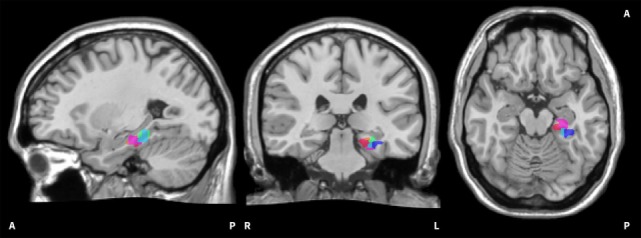
Figure 2.

Visualisation of on-line regions of interest (ROIs). The image shows all on-line ROIs from two exemplary subjects. The red/pink hues belong to the first subject, the blue/green hues to the second subject. It can be seen that the selected ROIs in the left parahippocampus are scattered to a certain extent. The ROIs are superimposed onto the Colin 27 average brain. Copyright© 1993–2009 Louis Collins, McConnell Brain Imaging Centre, Montreal Neurological Institute, McGill University.
