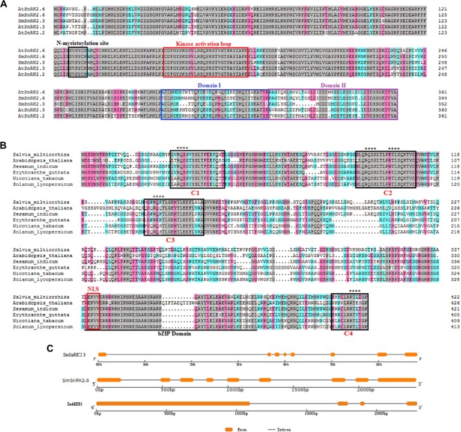
FIGURE 1.

Sequence analysis of SmSnRK2.3, SmSnRK2.6, and SmAREB1. (A) Multiple alignments of the deduced amino acid sequences of SmSnRK2.3 and SmSnRK2.6 with AtSnRK2.2, AtSnRK2.3, and AtSnRK2.6 from Arabidopsis thaliana (At). The regions of the N-myristoylation site, kinase activation loop, domain I, and domain II are boxed in black, red, blue, and violet, respectively. (B) Multiple alignment of the deduced amino acid sequence of SmAREB1 and the counterpart protein from other plants. The conserved C1, C2, C3, and C4 domains are boxed in black; the bZIP region is underlined; the NLS domain is boxed in red. The potential five phosphorylation recognition motifs (RXXS/T) are denoted with asterisks (∗). All multiple alignments were performed using DNAMAN. (C) The positions and length of exons and introns of SmSnRK2.3, SmSnRK2.6, and SmAREB1 are displayed schematically. Rounded rectangles indicate exons, while black lines indicate introns.
