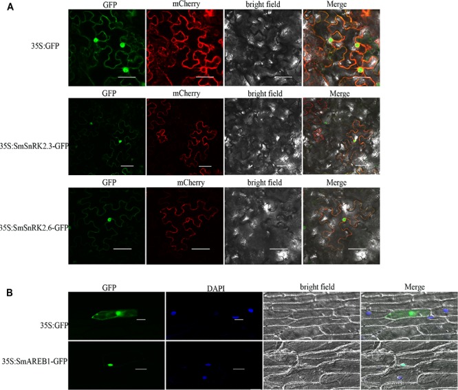
FIGURE 4.

Subcellular localization of SmSnRK2.3, SmSnRK2.6, and SmAREB1. (A) Subcellular localization of SmSnRK2.3 and SmSnRK2.6 in leaf epidermal cells of tobacco. GFP: green fluorescence; mCherry: red fluorescence of plasma membrane marker; Merge: merge of bright field and relevant fluorescences. Scale bar = 50 μm. (B) Subcellular localization of SmAREB1 in onion epidermal cells. GFP, green fluorescence; DAPI, fluorescence of DAPI nuclear dye; Merge, merge of bright field, GFP, and DAPI. Independent of (A) or (B), fluorescences of the empty vector pCAMBIA1301 were used as control. Scale bar = 50 μm.
