
Figure 4.
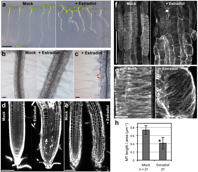
NEK6 overexpression disorganizes cortical microtubules and suppresses cell elongation. (a) Morphology of seedlings harboring estrogen-inducible NEK6. Seedlings were grown vertically for 8 days in the absence (Mock) or presence of estradiol (+Estradiol). The scale bar represents 1 cm. (b,c) Effect of NEK6 overexpression on epidermal cell morphology. Roots of seedlings grown for 8 days without (Mock) or with estradiol (+Estradiol) were observed under a microscope. Arrowheads indicate two root hairs in a trichoblast in the presence of estradiol. Scale bars represent 100 µm. (d,e) Middle-vertical section (d) and epidermal cells (e) of the root tip. The roots of seedlings grown for 8 days without (Mock) or with estradiol (+Estradiol) were stained with propidium iodide and observed under a confocal microscope. Scale bars represent 100 µm. (f) Effect of NEK6 overexpression on cortical microtubule arrays. The seedlings grown for 4 days without (Mock) or with estradiol (+Estradiol) were stained with anti-β-tubulin antibody. Fluorescence images show cortical microtubules in root epidermal cells in the elongation region. The scale bar represents 100 µm. (g) Enlarged view of cortical microtubule arrays in (f). The scale bar represents 10 µm. (h) Quantification of cortical microtubule density without (Mock) or with estradiol (+Estradiol). Data are displayed as averages ± SDs. The asterisk indicates significant difference from the value in mock-treated plants (t-test, P < 0.001).